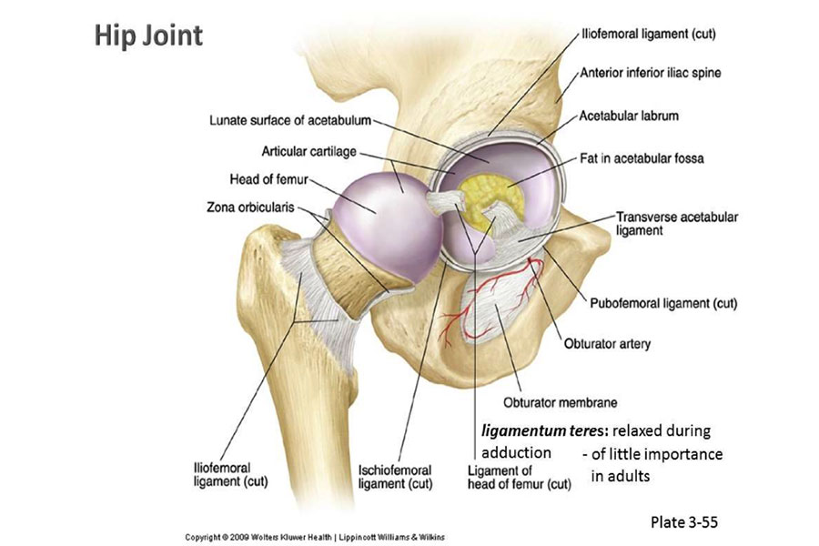
The hip is a joint comprised of a ball and socket. The acetabulum is the socket found on the pelvic bone; the femoral head on the upper end of the thighbone is the ball. Articular cartilage covers the ball and socket allowing for smooth movement with low friction. There is a generous blood supply within this area comprised of large arteries and veins allowing blood to flow to the femur and hip. Smaller blood vessels called capillaries also supply blood to the area within the ball and socket.
The hip joint is surrounded by tendons, ligaments and muscles that allow the femur to move as designed; yet keep it firmly stable within the socket. It takes a powerful force to dislodge the ball from the socket. This is called a dislocation.
For our civil newsletter and blog this month we are reviewing traumatic hip dislocations and delayed reduction. The blog topics for this month are:
- Hip Anatomy (3/2/15)
- Traumatic Dislocation (3/9/15)
- Avascular necrosis (3/16/15)
- Legal Implications (3/23/15)
Note: To see all posts in this topic, click here


